
Blank Diagram Of The Eye Diagrams Of Human Eye Printable ... 1.6 Many internal organs lie in membrane-lined body cavities: Human Anatomy and Physiology.
The middle layer, known as the vascular tunic or uvea, consists of the choroid, ciliary body, pigmented epithelium and iris.. The innermost is the retina, which ...Structure · Vision · Eye movement · Near response
30.07.2018 — Internal nervous coat is formed of retina.. The retina receives an inverted image of the objects seen.. These images are conducted to the brain ...7 Seiten
von CE Willoughby · 2010 · Zitiert von: 188 — The inner layer of the eye is the retina, a complex, layered structure of neurons that capture and process light.. The three transparent ...
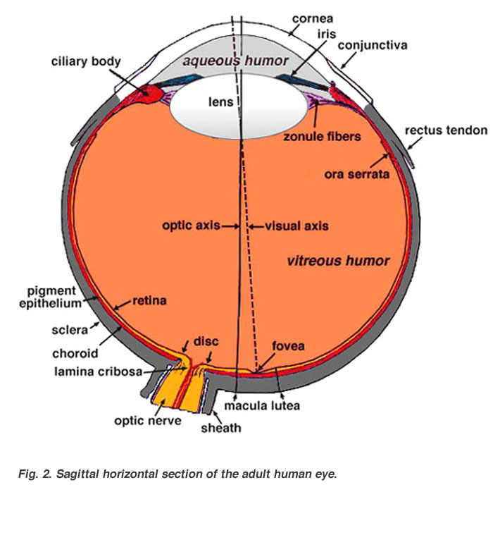
Anatomy of the Eye.. Schematic diagram of the human eye ... between the retina (the inner light-sensitive layer) and the sclera (the outer white eye wall).
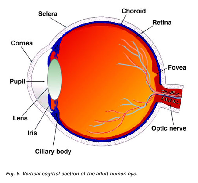
02.12.2019 — The main parts of the human eye are the cornea, iris, pupil, aqueous humor, lens, vitreous humor, retina, and optic nerve.. Light enters the eye ...
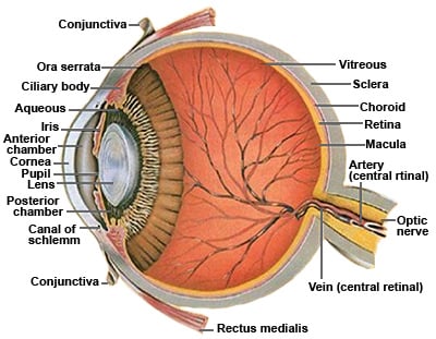
20.09.2019 — Illustration shows the right eyeball with a section removed to see the interior structures.. The retina.. Special cells called cones and rods are ...
11.09.2020 — Most of the eye is filled with a clear gel called the vitreous.. Light projects through your pupil and lens to the back of the eye.. The inside ...
... parts of our human eye illustration for descriptions of the eye anatomy; ... the retina — the light-sensitive inner lining of the back of the eye.
21.11.2016 — Layers of the eye; The inner part of the eyeball; How the eye works ..
Human eyes primarily consist of two globe-shaped structures, ...
A ring of muscular tissue, called the ciliary body, surrounds the lens and is connected to the lens by fine fibers, called zonules. how much does dog teeth cleaning
internal anatomy human
Together, the lens and the ...Facts & Orbit · Conjunctiva & Sclera · Chambers · Lens, Vitreous Cavity26.09.2018 — Retina is the innermost layer of the eyeball structure.. Retinal membrane can be imagined as the wall on which the images are projected.. The ...
The eye is our organ of sight.. The eye has a number of components which include but are not limited to the cornea, iris, pupil, lens, retina, macula, ...
The Internal Structure of the Eye · After the Iris and inside the Sclera is the Inner Structure of the eye.. This includes · Aqueous Humour · Vitreous Humour · Lens.
The internal components of an eye are: Lens: It is a transparent, biconvex, lens of an eye.. The lens is attached to the ciliary body by ligaments.. The lens ...13.08.2020
13.02.2020 — Internal parts of the eye ... Cornea: The cornea is the clear surface at the front of your eye, allowing light to enter the eye. cat teeth cleaning is it worth it
internal anatomy human body images
It directly ...
28.01.2021 — The cornea allows light to enter the eye.. As light passes through the eye the iris changes shape by expanding and letting more light through ...
19.03.2019 — The conjunctiva is a continuous, transparent mucous membrane, which lines the inner surface of the eyelids (and the outer surface of the eyes) ...
. why do all my bottom teeth hurt060951ff0b
